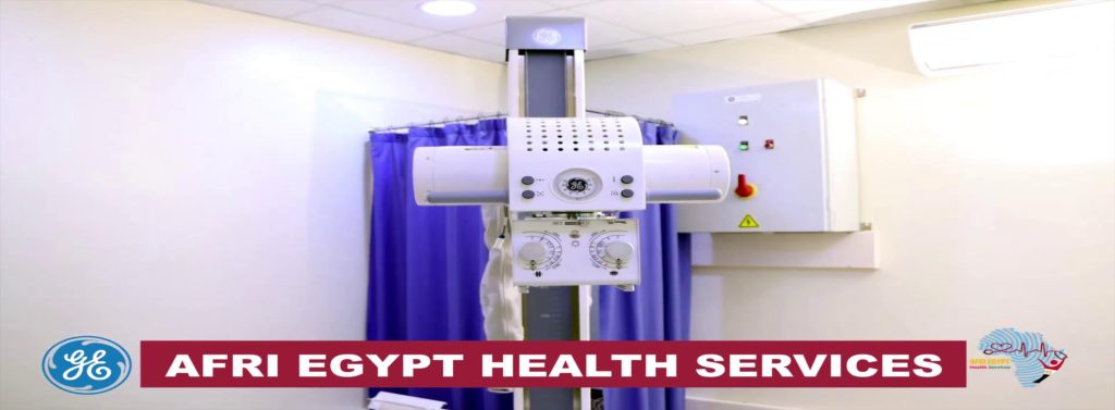
What is an X-ray?
An x-ray (radiograph) is a special image that uses radiation to create pictures of bones and other internal tissues such as your lungs and bowel. We use state of the art digital x-ray equipment, resulting in a reduced amount of radiation for excellent image quality.
What preparation is required?
A basic x-ray does not require any special preparation. Metal objects such as watches, keys, coins and jewellery will show up on the x-ray, affecting the images and therefore will need to be removed. You may be provided with a gown to wear instead of your own clothes, as some materials and prints will show up on the x-ray.
What happens during your X-ray?
You will be asked to stand or lie down in different positions that allow the best digital image of the body part of interest. During the x-ray, you will be asked to remain as still as possible or hold your breath in order to improve the quality of the images. As is the case with a regular photograph, any movement will appear blurry and may require the x-ray to be repeated. There are no after effects from a general x-ray. You will be able to go about your normal activities immediately following your x-ray.
How long will the X-ray take?
An x-ray will only take a few minutes for each body part. When the test is over, it may be necessary for you to wait while the images are reviewed by the Radiologist, to see if any further images are required.
What is the radiation dose?
Having an x-ray will expose your body to a very low level of radiation. Health experts feel that the risk to your health from this is very small and the low risk is outweighed by the benefits of your test. No radiation remains in your body after the test.
*If you are pregnant or think that you may be pregnant, tell your doctor and staff, as x-rays can potentially affect an unborn baby.
How much will the X-ray cost?
All x-rays are bulk billed (no out of pocket expense to the patient) to Medicare, provided that all Medicare eligibility requirements have been met.
Our Customer Service Team will be able to advise you of all costs involved with your x-ray including any out of pocket cost (if relevant).
When can I get my results?
Images obtained from your scan are digitally recorded. A subspecialty trained Radiologist interprets the images obtained and provides a report immediately.
Films are available for collection within one hour after finishing the study .
What Ultrasound services can we provide?
- General Ultrasound
- Echocardiogram
- Doppler studies
- Obstetric (Pregnancy)
- Thyroid
- Breast
- Musculoskeletal
- Vascular (veins and arteries)
- Pediatric (children)
- Interventional (Ultrasound-Guided Procedures)
General Ultrasound
Abdominal Ultrasound
Upper abdominal scans encompass the liver, pancreas, kidneys, gall bladder, spleen and aorta. If your doctor has requested a scan of these structures, you will be required to fast.
For adults, no food or liquid (other than sips of water) are permitted for 6 hours prior to the scheduled appointment time.
Children are required to fast for 4 hours. An early morning appointment is recommended.
Renal Ultrasound
Renal ultrasound scans assess the kidneys, bladder and prostate (in men).
You will be required to fast for 4-6 hours prior to your appointment.
We recommend that you finish drinking 1 litre of water an hour prior to the exam. Please do not empty your bladder after drinking the water.
Pelvic Ultrasound
Pelvic ultrasound scans assess the uterus and ovaries in women.
We recommend that you finish drinking 750mls of water an hour prior to the exam. Please do not empty your bladder after drinking the water.
Thyroid, Testicles, Breast and Small Part Ultrasounds
For these type of Ultrasound scans, no preparation is required.
Obstetric Ultrasound (Pregnancy)
Obstetric Ultrasound
We offers dating scans, Nuchal Translucency Screening Tests, Morphology and Third Trimester scans.
Musculoskeletal Ultrasound
Musculoskeletal Ultrasound
Our Radiology specialises in Musculoskeletal Ultrasound. This type of imaging uses sound waves to produce pictures of muscles, tendons, ligaments and joints throughout the body. It is used to help diagnose sprains, strains, tears and other soft tissue conditions. This is a safe, noninvasive and does not use ionizing radiation.
Vascular Ultrasound
Vascular Ultrasound
This type of imaging is a noninvasive ultrasound method (also called a duplex study) used to examine the circulation in the blood vessels of the body. Vascular Ultrasound can be used to evaluate arteries or veins in nearly any part of the body, including blood vessels in the neck, abdomen, arms and legs.
Certain Vascular Ultrasounds require specific preparation. These details will be provided to you at the time of booking.
Pediatric Ultrasound (Children)
Pediatric Ultrasound
We offers a range of Pediatric Ultrasounds including: Abdominal, Pediatric Spine, Pediatric Hip and Pediatric Head.
Interventional (Ultrasound-Guided Procedures)
What is Interventional Radiology?
Interventional radiology (abbreviated to IR or VIR for Vascular and Interventional Radiology) is a medical subspecialty of radiology that uses minimally invasive image guided procedures to diagnose and treat diseases in nearly every organ system including, but not limited to vascular, gastrointestinal, hepatobiliary, genitourinary, pulmonary, musculoskeletal, and the central nervous system.
The concept behind interventional radiology is to diagnose and treat patients using the least invasive techniques available to minimize risk, improve health outcomes and overall recovery time. Interventional radiology utilizes the latest technology in diagnostic imaging. The procedures can be performed using CT scan or ultrasound. The type of intervention requested will determine whether you are having a CT scan, ultrasound or fluoroscopy guided procedure.
Ultrasound policy
All ultrasound patients are advised to arrive 15 minutes prior to their appointment time with their doctors referral/request form.
An Ultrasound is a medical exam. It is therefore requested that all children under the age of 6 be supervised by another adult, other than the patient. To ensure the accuracy of the examination, children may not always be permitted in the room during the scan.
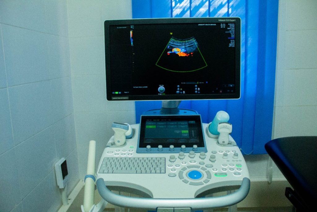
What is an Ultrasound?
An ultrasound scan is a procedure that uses high frequency sound waves to generate images of internal body structures.
Ultrasound has a wide range of applications. It is commonly used for obstetric imaging; small parts imaging, including the breast, neck and testes; abdominal and pelvic imaging; as well as musculoskeletal and vascular imaging.
UItrasound provides detailed, real time information without the use of radiation. The ability to see a moving image also makes ultrasound an excellent tool for guiding procedures such as needle biopsies and injections.
Images are generated with the use of a small hand held ultrasound transducer. High frequency sound waves, not audible to the human ear, are transmitted to the area of interest and the reflected signal is converted into an image. Specialist Radiologists then review these images for diagnostic purposes.
What preparation is required?
Preparation for an ultrasound will vary depending on the type of examination requested. Many examinations do not require any preparation. For your specific appointment, your preparation requirements will be outlined to you at the time of booking .
It is essential that your preparation needs are considered in conjunction with existing medical conditions such as diabetes or heart failure. If either of these conditions apply to you, or if you have any other concerns, please advise our Customer Service Team who will be able to provide you with personalised preparation information for your appointment.
You will need to bring your referral and any previous x-rays or scans with you to your appointment.
What happens during my Ultrasound?
Your Sonographer will collect you from the reception area and escort you through to the ultrasound room. Depending on your scan, you may be required to change into a gown. You will then be positioned accordingly for your scan.
A water-based gel will be applied to the relevant area to ensure that the transducer has adequate contact with your skin. The Sonographer will then use the transducer to obtain different views and record a series of images. This process is generally pain-free, however some pressure may need to be applied to improve the image. Your Sonographer may request that you also assist in certain scans, for example – holding your breath, which allows for improved views of certain structures.
How long will the Ultrasound take?
Your scan time will vary depending on the complexity of the requested examination. Most simple ultrasound scans take approximately 15-20 minutes, however more time is required for complex studies such as certain obstetric or vascular imaging.
When the scan is complete, it may be necessary for you to wait while our Radiologist reviews the obtained images. Occasionally further imaging may be required. Your Sonographer will advise you if this is the case.
What is the radiation dose?
Ultrasound uses sound waves only and does not use ionising radiation.
Ultrasound does not have any known risks, side effects or complications and is considered very safe.
How much will the Ultrasound cost?
The cost of the scan depends on a number of factors, including the type of scan that your doctor has requested. Please advise our friendly Customer Service Team if you are a pension or health care card holder.
If your doctor has requested more than one ultrasound, it may be necessary for these to be booked on separate days due to Medicare or different preparation requirements.
Our Customer Service Team will be able to advise you of all costs involved with your ultrasound including any out of pocket cost (if relevant).
When can I get my results?
Images obtained from your scan are digitally recorded. A subspecialty trained Radiologist interprets the images obtained and provides a report for you within 60 minutes.
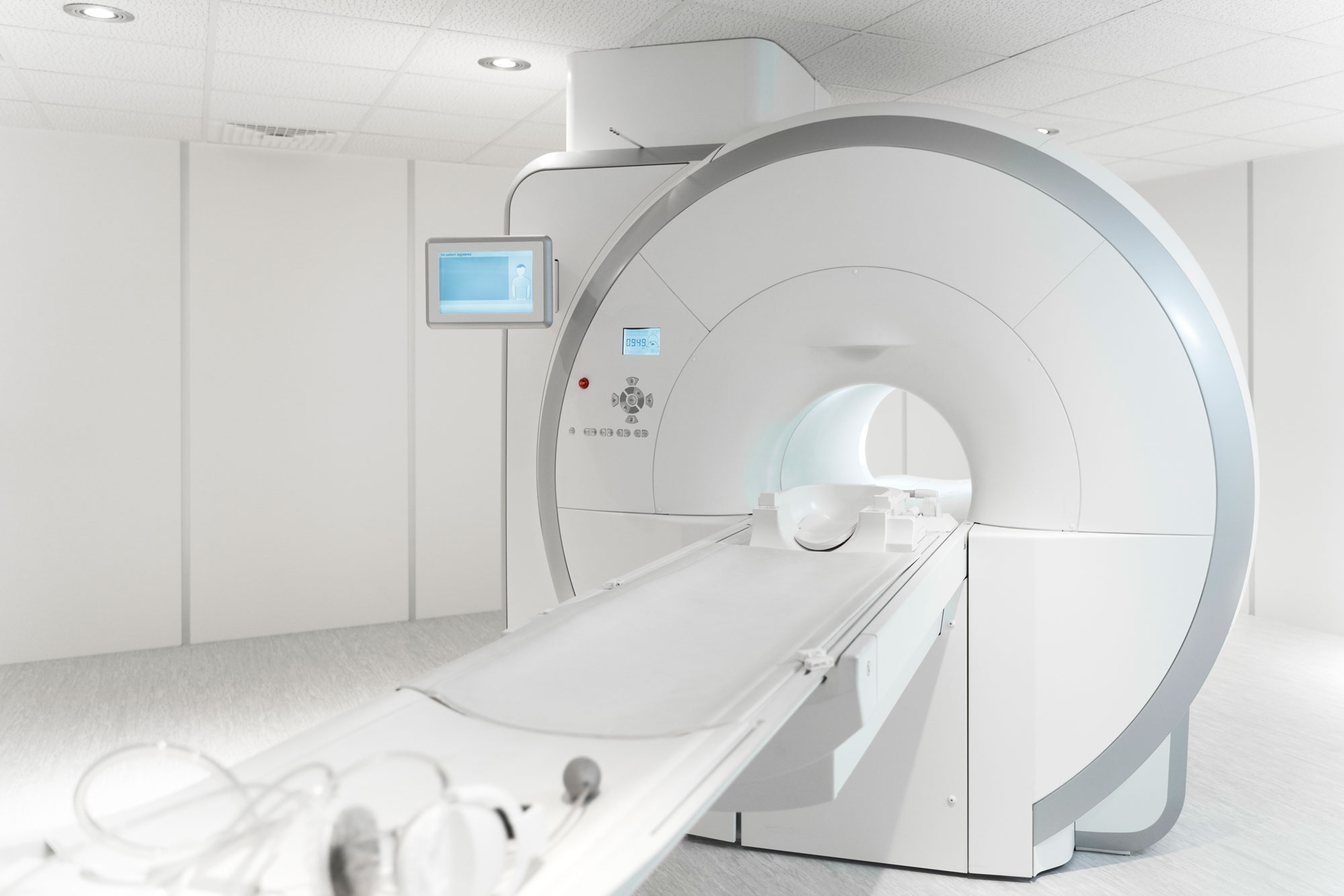
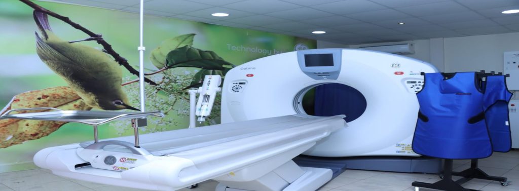
What is Computed Tomography?
Computed Tomography (CT) uses an x-ray machine and advanced computer programs to create two and three dimensional images of your body. CT is an essential imaging modality which utilizes the latest and most advanced medical technology to detect and assist with the diagnosis of many conditions.
What preparation is required?
Preparations, if required, can include being well hydrated, fasting for two hours prior to your scan or possible blood tests. Several CT examinations require preparation however one of our friendly Customer Service Team members will inform you of any specific preparation required for your CT scan at the time of your booking. Please see below an example of why specific preparation is required for certain body regions.
Abdominal and Pelvic CT
Depending on the clinical details provided by your referring doctor, some abdominal and pelvic CT scans may require you to arrive at our clinic 75 minutes prior to your actual scan time. This is to ensure that you have sufficient time to drink 1 litre of concentrated fluid (oral contrast) prior to the commencement of your scan. The purpose of oral contrast is to better demonstrate the stomach and bowel.
*If you are pregnant or think that you may be pregnant, please inform the Customer Service Team member at the time of booking your appointment as alternative medical imaging may need to be arranged.
What happens during your CT scan?
Metal objects such as watches, keys, coins and jewellery will show up on the CT scan, affecting the images and therefore will need to be removed. You may be provided with a gown to wear instead of your own clothes, as some materials and prints will show up on the CT scan.
You will be asked to lie on a long table that will move through the doughnut shaped CT scanner. The CT scanner is not an enclosed tunnel and for the majority of examinations your head will remain outside of the machine.
It is important to remain still and follow the specific instructions given to you at the start of your scan. You may be required to hold your breath for 5 -10 seconds at various stages throughout your examination to help with image quality.
How long will the CT scan take?
Timing of your scan will depend on the examination you are having. The actual scan may only take a matter of seconds, however more time is allowed for positioning you on the CT table, ensuring your comfort as well as providing you with instructions before beginning the examination.
Most examinations take no more than 5 – 20 minutes and you will be able to talk to CT staff at all times, should you have any questions.
What is the radiation dose?
All CT scans are performed using the lowest radiation dose possible. Every CT scanner utilizes advanced dose saving technology to optimize low dose scanning techniques.
How much will the CT scan cost?
Our Customer Service Team will be able to advise you of all costs involved with your CT scan including any out of pocket cost before your scan. Many CT scans are entitled to be bulk-billed, provided that all Medicare eligibility requirements have been met.
When can I get my results?
Images obtained from your scan are digitally recorded. A subspecialty trained Radiologist interprets the images obtained and provides a report immediately.
Films are available for collection within one hour after finishing the study .
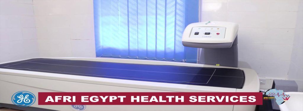
What is Bone Mineral Densitometry?
Bone Mineral Densitometry (BMD) is a scan which uses low energy x-rays to measure the density of your bone. It is used to diagnose osteoporosis and assess your fracture risk. It is also used to monitor your bone density which is especially useful if you are on treatment for osteoporosis. The usual regions scanned are your lower spine and hips although on occasions other areas such as the forearm may need to be scanned.
What preparation is required?
There is no special preparation required for a bone mineral densitometry scan.
Where possible, it is best to wear clothing with no metallic objects such as zips or metal buttons as this can affect the results of the scan. However if required, a gown will be provided before scanning.
It is recommended to bring along any previous bone mineral densitometry results that you have as any changes in your bone density can be assessed by comparing to your previous results.
What happens during your Bone Mineral Densitometry scan?
Before the commencement of your scan, your height, weight and some medical history will be obtained. You will be asked to lie on a scanning bed. Usually the lower spine and both hips are scanned however this may vary depending on your clinical circumstances.
How long will your Bone Mineral Densitometry scan take?
The total duration of your appointment will be approximately 20 minutes however the scanning time is around 5 minutes.
What is the radiation dose?
The radiation dose from a BMD scan is very low and is about one tenth of a dose from a chest x-ray.
How much will the Bone Mineral Densitometry scan cost?
The cost of a bone mineral densitometry scan depends on whether you meet Medicare criteria for a rebate. Please discuss your eligibility for a rebate with your referring doctor prior to your appointment .
Our Customer Service Team will be able to advise you of all costs involved with your BMD including any out of pocket cost.
When can I get my results?
Images obtained from your scan are digitally recorded. A subspecialty trained Radiologist interprets the images obtained and provides a report immediately.
Films are available for collection within one hour after finishing the study .
What is mammography?
A diagnostic mammogram is an x-ray examination of the breasts. This examination is performed when your doctor suspects unusual signs or symptoms in one or both breasts, for example a lump, tenderness, and nipple discharge or skin changes.
A 3D Mammogram captures a series of thin ‘sections (around 1mm thick) through the breast providing greater detail. 3D Mammography can demonstrate early invasive breast cancers more clearly than 2D Mammography alone. 3D Mammograms may be more valuable for those with dense breast tissue or implants.
What preparation is required?
No perfume, deodorant, lotions (e.g. moisture cream) or talcum powder as this can show up on the x-ray and impact the accuracy of the image.
Wearing a two piece outfit that allows you to continue to wear the bottom section (e.g. trousers or skirt) but for ease of examination allows the top section (shirt) to be removed. This will assist with providing privacy and warmth.
If you experience significant breast discomfort during your menstrual period, it may be best not to schedule your mammogram during this time. One week after you period is best, unless the examination is urgent.
Eat and drink normally and continue to take you usual medications.
It is very important to bring previous films to your appointment – even if your previous Mammogram was done by us. This is to support a thorough analysis of your examination.
What happens during my Mammogram?
Mammograms are performed by a radiographer who has received specialist training in the field of mammography. Each breast will be positioned and compressed between two special plates by the X-ray machine for a few seconds while images are taken. The mammogram is then read and interpreted by a radiologist who will provide your referring doctor with a report of your examination.
While the examination may be uncomfortable, it will last only a few seconds.
What are the benefits of having a Mammogram?
Multiple scientific studies have provided plenty of evidence that early diagnosis and treatment of breast cancer can save lives. Early detection increases the likelihood of a cancer being successfully treated and often allows for greater treatment options.
It is important to note that mammography does not detect all breast cancers and a normal mammogram does not mean that the lump can be ignored. In some circumstances, other diagnostic tests such as a breast ultrasound and fine need biopsy may be necessary to find out the cause of the lump.
Are there any side effects?
After effects are rare, however, you may experience breast tenderness or bruising. Should you have any concerns please let us or your doctor know.
How long does a Mammogram take?
A standard mammography takes between 10-20 minutes. Some extra views may be performed which take longer.
If you have breast implants, please advise our practice prior to your appointment so that a longer appointment is scheduled.
How much will the Mammogram cost?
Depending on your clinical history there may be a fee for your examination. Please ask one of our team at the time of your booking for more information in relation to our specific circumstances.
When can I get my results?
Images obtained from your scan are digitally recorded. A subspecialty trained Radiologist interprets the images obtained and provides a report immediately.
Films are available for collection within one hour after finishing the study .
x-ray
An x-ray (radiograph) is a special image that uses radiation to create pictures of bones and other internal tissues such as your lungs and bowel. We use state of the art digital x-ray equipment, resulting in a reduced amount of radiation for excellent image quality.
Ultrasound
An ultrasound scan is a procedure that uses high frequency sound waves to generate images of internal body structures.Ultrasound has a wide range of applications. It is commonly used for obstetric imaging; small parts imaging, including the breast, neck and testes; abdominal and pelvic imaging; as well as musculoskeletal and vascular imaging.
Computed Tomography (CT)
Computed Tomography (CT) uses an x-ray machine and advanced computer programs to create two and three dimensional images of your body. CT is an essential imaging modality which utilizes the latest and most advanced medical technology to detect and assist with the diagnosis of many conditions.
Bone Mineral Densitometry
Bone Mineral Densitometry (BMD) is a scan which uses low energy x-rays to measure the density of your bone. It is used to diagnose osteoporosis and assess your fracture risk. It is also used to monitor your bone density which is especially useful if you are on treatment for osteoporosis. The usual regions scanned are your lower spine and hips although on occasions other areas such as the forearm may need to be scanned.
Mammography
A diagnostic mammogram is an x-ray examination of the breasts. This examination is performed when your doctor suspects unusual signs or symptoms in one or both breasts, for example a lump, tenderness, and nipple discharge or skin changes.
A 3D Mammogram captures a series of thin ‘sections (around 1mm thick) through the breast providing greater detail. 3D Mammography can demonstrate early invasive breast cancers more clearly than 2D Mammography alone. 3D Mammograms may be more valuable for those with dense breast tissue or implants.